
Electron Probe Microanalyzer
Instrument: JXA - 8230
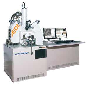
The JXA-8230 Electron Probe Microanalyzer measures the energy and wavelengths of characteristic x-rays by sending a beam of electrons to the sample. It also has the ability to perform qualitative and full quantitative analysis.
Specifications
Element Measurement Range: B(5) to U(92)
Resolution (SEI): 6nm
Magnification : 40x to 300.000x
Acceleration Voltage: 0.2 to 30 kV
Image Mode: SEI (Scanning Electron Microscope)
BEI (Backscattered Electron Imaging)
OMI (Optical Microscope Image in Reflection and Transmission Mode)
XRAY (X-Ray mapping for elements B(5) to U(92))
Instrument: JXA - 8230
The JXA-8230 Electron Probe Microanalyzer measures the energy and wavelengths of characteristic x-rays by sending a beam of electrons to the sample. It also has the ability to perform qualitative and full quantitative analysis.
Specifications
Element Measurement Range: B(5) to U(92)
Resolution (SEI): 6nm
Magnification : 40x to 300.000x
Acceleration Voltage: 0.2 to 30 kV
Image Mode: SEI (Scanning Electron Microscope)
BEI (Backscattered Electron Imaging)
OMI (Optical Microscope Image in Reflection and Transmission Mode)
XRAY (X-Ray mapping for elements B(5) to U(92))